1.3 Optical Images
The scanner collects good quality optical images that have consistent lighting and because they are line-scan images, they do not suffer from optical distortions. Medium resolution images are usually included in all scan section folders (typically optical.tif), and optional high-resolution images are often included by operators elsewhere. High resolution images are typically supplied as both 8-bit and 16-bit images, and are usually hundreds of megabytes in size.
Although the brightness of the lighting in the scanner is adjustable, images sometimes need to be brightened and/or have the contrast adjusted. This can be performed in desktop publishing software (e.g. Adobe Photoshop, Corel Photo-Draw), but it is easiest to use the open-source scientific image analysis software imageJ (NIH, USA).
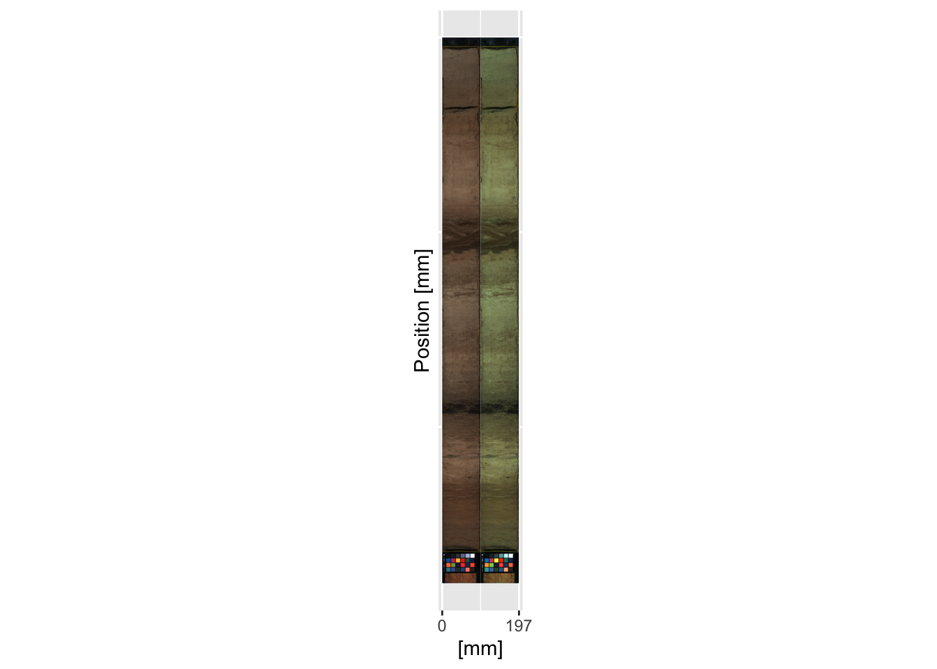
By including an appropriate colour reference card in the scan the image can be calibrated, although it is often desirable to increase the contrast and gamma to elucidate features of the core. The photograph is always of the entire length of the bed scanned, rather than cropped to the limits of an individual scan section. If multiple scan sections are placed on the bed and are scanned together, it will include all of the scanned sections. The image needs to be cropped using the metadata for the relevant scan section; this process is covered in later chapters.
In this case, the colour reference information for the card in the image is given in the file greywhitebalancecolourcard.csv. The plugin for imageJ, ijp-color, can be used to perform colour calibration on the images. In this case a linear - no intercept model has been used. to generate the image files image0.tif. The image below shows the uncorrected image (left hand side) and the colour corrected image (right hand side).